

Article Sidebar
Main Article Content
Abstract
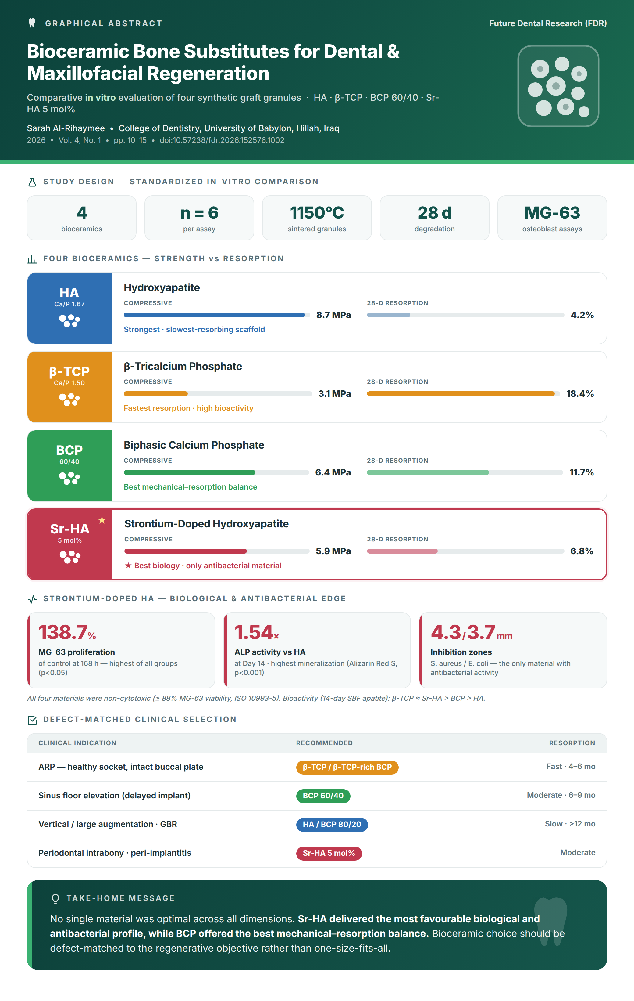
Background: Bioceramic bone substitutes are central to contemporary dental bone regeneration, but the relative physicochemical, mechanical, and biological merits of stoichiometric hydroxyapatite (HA), β-tricalcium phosphate (β-TCP), biphasic calcium phosphate (BCP), and strontium-doped HA (Sr-HA) remain insufficiently characterized within a single, controlled in vitro framework. This study compared four laboratory-synthesized bioceramic granules under identical processing and testing conditions.
Methods: HA, β-TCP, BCP (60/40 HA: β-TCP), and 5 mol% Sr-HA powders were synthesized by aqueous chemical precipitation, calcined at 900 °C, and sintered as 5 × 5 × 5 mm granules at 1150 °C. Phase composition was determined by XRD; functional groups by FTIR; morphology and elemental ratios by SEM-EDS; surface area by BET. Mechanical strength and porosity were quantified; in vitro bioactivity was assessed by 14-day SBF immersion; degradation by 28-day Tris-HCl weight loss. MG-63 cell viability (MTT, ISO 10993-5), ALP activity, and Alizarin Red S mineralization were evaluated. Antibacterial activity was tested against S. aureus ATCC 25923 and E. coli ATCC 25922 by agar diffusion. Data (n = 6) were analyzed by one-way ANOVA and Tukey's test (α = 0.05).
Results: XRD confirmed phase-pure compositions. Compressive strength differed significantly (p < 0.001): HA 8.7 ± 0.6 MPa > BCP 6.4 ± 0.5 > Sr-HA 5.9 ± 0.5 > β-TCP 3.1 ± 0.4 MPa. Twenty-eight-day weight loss was highest for β-TCP (18.4 ± 1.6%) and lowest for HA (4.2 ± 0.5%). All materials were non-cytotoxic (≥88% viability). Sr-HA produced the highest MG-63 proliferation at 168 h (138.7 ± 7.4%; p < 0.05), ALP activity (1.54-fold over HA at Day 14; p < 0.001), mineralization (p < 0.001), and the only measurable antibacterial inhibition (S. aureus 4.3 ± 0.6 mm; E. coli 3.7 ± 0.5 mm).
Conclusion: No single material was optimal across all dimensions. Sr-HA provided the most favorable biological profile; BCP offered the best mechanical-resorption balance. A defect-matched clinical decision framework is proposed.
Keywords
Article Details
Copyright (c) 2026 Sarah alrihaymee (Author)

This work is licensed under a Creative Commons Attribution 4.0 International License.
How to Cite
References
- Tan WL, Wong TL, Wong MC, Lang NP. A systematic review of post‐extractional alveolar hard and soft tissue dimensional changes in humans. Clin Oral Implants Res. 2012;23(s5):1-21. doi: 10.1111/j.600-0501.2011.02375.x.
- Adams RJ. Is there clinical evidence to support alveolar ridge preservation over extraction alone? A review of recent literature and case reports of late graft failure. Br Dent J. 2022;233(6):469-74. doi: 10.1038/s41415-022-4967-2.
- Markets and Markets. Dental Bone Graft Substitute Market. 2026. https://www.marketsandmarkets.com/PressReleases/dental-bone-graft-substitutes.asp.
- Dorozhkin SV. Bioceramics of calcium orthophosphates. Biomaterials. 2010;31(7):1465-85. doi: 10.1016/j.biomaterials.2009.11.050.
- Stevens MM. Biomaterials for bone tissue engineering. Mater Today. 2008;11(5):18-25. doi: 10.1016/S369-7021(08)70086-5.
- Fendi F, Abdullah B, Suryani S, Usman AN, Tahir D. Development and application of hydroxyapatite-based scaffolds for bone tissue regeneration: A systematic literature review. Bone. 2024;183:117075. doi: 10.1016/j.bone.2024.
- Boskey AL. Bone composition: relationship to bone fragility and antiosteoporotic drug effects. BoneKEy Rep. 2013;2:447. doi: https://doi.org/10.1038/bonekey.2013.181.
- Horowitz RA, Mazor Z, Foitzik C, Prasad H, Rohrer M, Palti A. β-tricalcium phosphate as bone substitute material: properties and clinical applications. J Osseointegration. 2010;2(2):61-8. doi: 10.23805/jo.2010.02.02.04.
- Somngam C, Samartkit S, Kanchanasurakit S, Strietzel FP, Khongkhunthian P. New bone formation of biphasic calcium phosphate bone substitute material: a systematic review and network meta-analysis of randomized controlled trials (RCTs). Int J Implant Dent. 2025;11:47. doi: 10.1186/s40729-025-00636-4.
- Liu X, Huang H, Zhang J, Sun T, Zhang W, Li Z. Recent advance of strontium functionalized in biomaterials for bone regeneration. Bioengineering. 2023;10(4):414. doi: 10.3390/bioengineering10040414.
- Landi E, Tampieri A, Celotti G, Sprio S, Sandri M, Logroscino G. Sr-substituted hydroxyapatites for osteoporotic bone replacement. Acta Biomater. 2007;3(6):961-9. doi: 10.1016/j.actbio.2007.05.006.
- Tsai S-W, Hsu Y-W, Pan W-L, Hsu F-Y. The effect of strontium-substituted hydroxyapatite nanofibrous matrix on osteoblast proliferation and differentiation. Membranes. 2021;11(8):624. doi: 10.3390/membranes11080624.
- Cheng D, Ding R, Jin X, Lu Y, Bao W, Zhao Y, et al. Strontium ion-functionalized nano-hydroxyapatite/chitosan composite microspheres promote osteogenesis and angiogenesis for bone regeneration. ACS Appl Mater Interfaces. 2023;15(16):19951-65. doi: 10.1021/acsami.3c00655.
- Badea MA, Balas M, Popa M, Borcan T, Bunea A-C, Predoi D, et al. Biological response of human gingival fibroblasts to zinc-doped hydroxyapatite designed for dental applications—an in vitro study. Materials. 2023;16(11):4145. doi: 10.3390/ma16114145.
- Balamurugan A, Rebelo A, Lemos A, Rocha J, Ventura J, Ferreira J. Suitability evaluation of sol–gel derived Si-substituted hydroxyapatite for dental and maxillofacial applications through in vitro osteoblasts response. Dent Mater. 2008;24(10):1374-80. doi: 10.016/j.dental.2008.02.017.
- Bandyopadhyay A, Bernard S, Xue W, Bose S. Calcium phosphate-based resorbable ceramics: influence of MgO, ZnO, and SiO2 dopants. J Am Ceram Soc. 2006;89(9):2675-88. doi: 10.1111/j.1551-2916.2006.01207.x.
- Frasnelli M, Sglavo VM. Effect of Mg2+ doping on beta–alpha phase transition in tricalcium phosphate (TCP) bioceramics. Acta Biomater. 2016;33:283-9. doi: 10.1016/j.actbio.2016.01.015.
- Kokubo T, Takadama H. How useful is SBF in predicting in vivo bone bioactivity? Biomaterials. 2006;27(15):2907-15. doi: 10.1016/j.biomaterials.2006.01.017.
- Kokubo T, Yamaguchi S. Simulated body fluid and the novel bioactive materials derived from it. J Biomed Mater Res A. 2019;107(5):968-77. doi: 10.1002/jbm.a.36620.
- Champion E. Sintering of calcium phosphate bioceramics. Acta Biomater. 2013;9(4):5855-75. doi: 10.1016/j.actbio.2012.11.029.
- Frasnelli M, Cristofaro F, Sglavo VM, Dirè S, Callone E, Ceccato R, et al. Synthesis and characterization of strontium-substituted hydroxyapatite nanoparticles for bone regeneration. Mater Sci Eng C. 2017;71:653-62. doi: 10.1016/j.msec.2016.10.047.
- Capuccini C, Torricelli P, Sima F, Boanini E, Ristoscu C, Bracci B, et al. Strontium-substituted hydroxyapatite coatings synthesized by pulsed-laser deposition: in vitro osteoblast and osteoclast response. Acta Biomater. 2008;4(6):1885-93. doi: 10.016/j.actbio.2008.05.005.
- Bohner M, Santoni BLG, Döbelin N. β-tricalcium phosphate for bone substitution: Synthesis and properties. Acta Biomater. 2020;113:23-41. doi: 10.1016/j.actbio.2020.06.022.
- Liang H, Wang Y, Chen S, Liu Y, Liu Z, Bai J. Nano-hydroxyapatite bone scaffolds with different porous structures processed by digital light processing 3D printing. Int J Bioprint. 2022;8(1):502. doi: 10.18063/ijb.v8i1.502.
- Diez-Escudero A, Espanol M, Beats S, Ginebra M-P. In vitro degradation of calcium phosphates: Effect of multiscale porosity, textural properties and composition. Acta Biomater. 2017;60:81-92. doi: 10.1016/j.actbio.2017.07.033.
- Garrido CA, Lobo SE, Turíbio FM, LeGeros RZ. Biphasic calcium phosphate bioceramics for orthopaedic reconstructions: clinical outcomes. Int J Biomater. 2011;2011:129727. doi: 10.1155/2011/.
- Ran L, Liu L, Gao J, Pan Y, Ramalingam M, Du X, et al. Strontium-doped hydroxyapatite and its role in osteogenesis and angiogenesis. Int J Dev Biol. 2023;67(4):137-46. doi: 10.1387/ijdb.230091lc.
- Yang L, Perez-Amodio S, Barrère-de Groot FYF, Everts V, van Blitterswijk CA, Habibovic P. The effects of inorganic additives to calcium phosphate on in vitro behavior of osteoblasts and osteoclasts. Biomaterials. 2010;31(11):2976-89. doi: 10.1016/j.biomaterials.2010.01.002.
- Predoi D, Iconaru SL, Deniaud A, Chevallet M, Michaud-Soret I, Buton N, et al. Textural, structural and biological evaluation of hydroxyapatite doped with zinc at low concentrations. Materials. 2017;10(3):229. doi: 10.3390/ma10030229.
- Mocanu A, Cadar O, Frangopol PT, Petean I, Tomoaia G, Paltinean G-A, et al. Ion release from hydroxyapatite and substituted hydroxyapatites in different immersion liquids: In vitro experiments and theoretical modelling study. R Soc Open Sci. 2021;8(1):201785. doi: 10.1098/rsos.
References
Tan WL, Wong TL, Wong MC, Lang NP. A systematic review of post‐extractional alveolar hard and soft tissue dimensional changes in humans. Clin Oral Implants Res. 2012;23(s5):1-21. doi: 10.1111/j.600-0501.2011.02375.x.
Adams RJ. Is there clinical evidence to support alveolar ridge preservation over extraction alone? A review of recent literature and case reports of late graft failure. Br Dent J. 2022;233(6):469-74. doi: 10.1038/s41415-022-4967-2.
Markets and Markets. Dental Bone Graft Substitute Market. 2026. https://www.marketsandmarkets.com/PressReleases/dental-bone-graft-substitutes.asp.
Dorozhkin SV. Bioceramics of calcium orthophosphates. Biomaterials. 2010;31(7):1465-85. doi: 10.1016/j.biomaterials.2009.11.050.
Stevens MM. Biomaterials for bone tissue engineering. Mater Today. 2008;11(5):18-25. doi: 10.1016/S369-7021(08)70086-5.
Fendi F, Abdullah B, Suryani S, Usman AN, Tahir D. Development and application of hydroxyapatite-based scaffolds for bone tissue regeneration: A systematic literature review. Bone. 2024;183:117075. doi: 10.1016/j.bone.2024.
Boskey AL. Bone composition: relationship to bone fragility and antiosteoporotic drug effects. BoneKEy Rep. 2013;2:447. doi: https://doi.org/10.1038/bonekey.2013.181.
Horowitz RA, Mazor Z, Foitzik C, Prasad H, Rohrer M, Palti A. β-tricalcium phosphate as bone substitute material: properties and clinical applications. J Osseointegration. 2010;2(2):61-8. doi: 10.23805/jo.2010.02.02.04.
Somngam C, Samartkit S, Kanchanasurakit S, Strietzel FP, Khongkhunthian P. New bone formation of biphasic calcium phosphate bone substitute material: a systematic review and network meta-analysis of randomized controlled trials (RCTs). Int J Implant Dent. 2025;11:47. doi: 10.1186/s40729-025-00636-4.
Liu X, Huang H, Zhang J, Sun T, Zhang W, Li Z. Recent advance of strontium functionalized in biomaterials for bone regeneration. Bioengineering. 2023;10(4):414. doi: 10.3390/bioengineering10040414.
Landi E, Tampieri A, Celotti G, Sprio S, Sandri M, Logroscino G. Sr-substituted hydroxyapatites for osteoporotic bone replacement. Acta Biomater. 2007;3(6):961-9. doi: 10.1016/j.actbio.2007.05.006.
Tsai S-W, Hsu Y-W, Pan W-L, Hsu F-Y. The effect of strontium-substituted hydroxyapatite nanofibrous matrix on osteoblast proliferation and differentiation. Membranes. 2021;11(8):624. doi: 10.3390/membranes11080624.
Cheng D, Ding R, Jin X, Lu Y, Bao W, Zhao Y, et al. Strontium ion-functionalized nano-hydroxyapatite/chitosan composite microspheres promote osteogenesis and angiogenesis for bone regeneration. ACS Appl Mater Interfaces. 2023;15(16):19951-65. doi: 10.1021/acsami.3c00655.
Badea MA, Balas M, Popa M, Borcan T, Bunea A-C, Predoi D, et al. Biological response of human gingival fibroblasts to zinc-doped hydroxyapatite designed for dental applications—an in vitro study. Materials. 2023;16(11):4145. doi: 10.3390/ma16114145.
Balamurugan A, Rebelo A, Lemos A, Rocha J, Ventura J, Ferreira J. Suitability evaluation of sol–gel derived Si-substituted hydroxyapatite for dental and maxillofacial applications through in vitro osteoblasts response. Dent Mater. 2008;24(10):1374-80. doi: 10.016/j.dental.2008.02.017.
Bandyopadhyay A, Bernard S, Xue W, Bose S. Calcium phosphate-based resorbable ceramics: influence of MgO, ZnO, and SiO2 dopants. J Am Ceram Soc. 2006;89(9):2675-88. doi: 10.1111/j.1551-2916.2006.01207.x.
Frasnelli M, Sglavo VM. Effect of Mg2+ doping on beta–alpha phase transition in tricalcium phosphate (TCP) bioceramics. Acta Biomater. 2016;33:283-9. doi: 10.1016/j.actbio.2016.01.015.
Kokubo T, Takadama H. How useful is SBF in predicting in vivo bone bioactivity? Biomaterials. 2006;27(15):2907-15. doi: 10.1016/j.biomaterials.2006.01.017.
Kokubo T, Yamaguchi S. Simulated body fluid and the novel bioactive materials derived from it. J Biomed Mater Res A. 2019;107(5):968-77. doi: 10.1002/jbm.a.36620.
Champion E. Sintering of calcium phosphate bioceramics. Acta Biomater. 2013;9(4):5855-75. doi: 10.1016/j.actbio.2012.11.029.
Frasnelli M, Cristofaro F, Sglavo VM, Dirè S, Callone E, Ceccato R, et al. Synthesis and characterization of strontium-substituted hydroxyapatite nanoparticles for bone regeneration. Mater Sci Eng C. 2017;71:653-62. doi: 10.1016/j.msec.2016.10.047.
Capuccini C, Torricelli P, Sima F, Boanini E, Ristoscu C, Bracci B, et al. Strontium-substituted hydroxyapatite coatings synthesized by pulsed-laser deposition: in vitro osteoblast and osteoclast response. Acta Biomater. 2008;4(6):1885-93. doi: 10.016/j.actbio.2008.05.005.
Bohner M, Santoni BLG, Döbelin N. β-tricalcium phosphate for bone substitution: Synthesis and properties. Acta Biomater. 2020;113:23-41. doi: 10.1016/j.actbio.2020.06.022.
Liang H, Wang Y, Chen S, Liu Y, Liu Z, Bai J. Nano-hydroxyapatite bone scaffolds with different porous structures processed by digital light processing 3D printing. Int J Bioprint. 2022;8(1):502. doi: 10.18063/ijb.v8i1.502.
Diez-Escudero A, Espanol M, Beats S, Ginebra M-P. In vitro degradation of calcium phosphates: Effect of multiscale porosity, textural properties and composition. Acta Biomater. 2017;60:81-92. doi: 10.1016/j.actbio.2017.07.033.
Garrido CA, Lobo SE, Turíbio FM, LeGeros RZ. Biphasic calcium phosphate bioceramics for orthopaedic reconstructions: clinical outcomes. Int J Biomater. 2011;2011:129727. doi: 10.1155/2011/.
Ran L, Liu L, Gao J, Pan Y, Ramalingam M, Du X, et al. Strontium-doped hydroxyapatite and its role in osteogenesis and angiogenesis. Int J Dev Biol. 2023;67(4):137-46. doi: 10.1387/ijdb.230091lc.
Yang L, Perez-Amodio S, Barrère-de Groot FYF, Everts V, van Blitterswijk CA, Habibovic P. The effects of inorganic additives to calcium phosphate on in vitro behavior of osteoblasts and osteoclasts. Biomaterials. 2010;31(11):2976-89. doi: 10.1016/j.biomaterials.2010.01.002.
Predoi D, Iconaru SL, Deniaud A, Chevallet M, Michaud-Soret I, Buton N, et al. Textural, structural and biological evaluation of hydroxyapatite doped with zinc at low concentrations. Materials. 2017;10(3):229. doi: 10.3390/ma10030229.
Mocanu A, Cadar O, Frangopol PT, Petean I, Tomoaia G, Paltinean G-A, et al. Ion release from hydroxyapatite and substituted hydroxyapatites in different immersion liquids: In vitro experiments and theoretical modelling study. R Soc Open Sci. 2021;8(1):201785. doi: 10.1098/rsos.